Animal Cell Diagram with Labels Explained
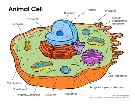
Understanding the Animal Cell Diagram with Labels
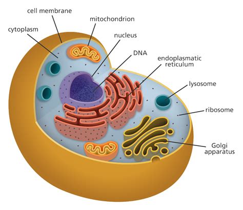
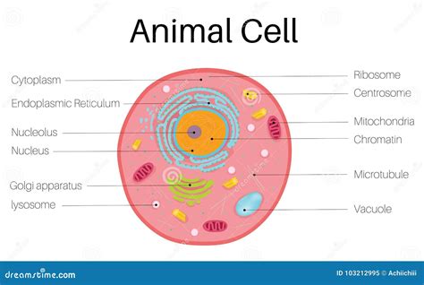
When it comes to understanding the intricate structures of living organisms, cell biology plays a crucial role. The animal cell, being a fundamental unit of life, is a fascinating entity that consists of various organelles, each with distinct functions. In this post, we will delve into the world of animal cells, exploring the different components of an animal cell diagram with labels, and explaining the roles they play.
Components of an Animal Cell Diagram
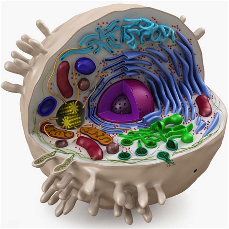
An animal cell diagram is a visual representation of the various organelles present within an animal cell. Here are the key components:
| Organelle | Function |
|---|---|
| Plasma Membrane | The outermost layer of the cell, responsible for regulating the movement of materials in and out of the cell. |
| Cytoplasm | A jelly-like substance that fills the cell, providing a medium for organelles to function and allowing for cell growth and division. |
| Nucleus | The control center of the cell, containing the genetic material (DNA) and regulating cell growth, division, and function. |
| Mitochondria | Responsible for generating energy for the cell through the process of cellular respiration. |
| Endoplasmic Reticulum (ER) | A network of membranous tubules and cisternae responsible for protein synthesis, transport, and storage. |
| Ribosomes | Small organelles found throughout the cytoplasm, responsible for protein synthesis. |
| Lysosomes | Membrane-bound sacs containing digestive enzymes, responsible for cellular digestion and recycling of waste materials. |
| Golgi Apparatus | A complex organelle responsible for protein modification, sorting, and packaging for transport out of the cell. |
| Cytoskeleton | A network of protein filaments providing structural support, shape, and movement to the cell. |
| Peroxisomes | Small organelles involved in the breakdown of fatty acids and amino acids. |
| Centrioles | Involved in the formation of cilia, flagella, and the spindle fibers that separate chromosomes during cell division. |
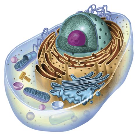
Exploring the Functions of Each Organelle
Now that we have identified the various components of an animal cell diagram, let’s take a closer look at the functions of each organelle:
- Plasma Membrane: The plasma membrane is semi-permeable, allowing certain substances to pass through while restricting others. This regulation is crucial for maintaining cellular homeostasis.
- Cytoplasm: The cytoplasm provides a medium for organelles to function and allows for cell growth and division. It also plays a role in cell signaling and the transport of molecules.
- Nucleus: The nucleus contains the genetic material (DNA) and regulates cell growth, division, and function. It is often referred to as the “control center” of the cell.
- Mitochondria: Mitochondria are responsible for generating energy for the cell through the process of cellular respiration. They are often referred to as the “powerhouses” of the cell.
- Endoplasmic Reticulum (ER): The ER is a network of membranous tubules and cisternae responsible for protein synthesis, transport, and storage. It is divided into two types: rough ER (with ribosomes) and smooth ER (without ribosomes).
- Ribosomes: Ribosomes are small organelles found throughout the cytoplasm, responsible for protein synthesis. They read messenger RNA (mRNA) sequences and assemble amino acids into proteins.
- Lysosomes: Lysosomes are membrane-bound sacs containing digestive enzymes, responsible for cellular digestion and recycling of waste materials.
- Golgi Apparatus: The Golgi apparatus is a complex organelle responsible for protein modification, sorting, and packaging for transport out of the cell.
- Cytoskeleton: The cytoskeleton provides structural support, shape, and movement to the cell. It is composed of three main components: microtubules, microfilaments, and intermediate filaments.
- Peroxisomes: Peroxisomes are small organelles involved in the breakdown of fatty acids and amino acids.
- Centrioles: Centrioles are involved in the formation of cilia, flagella, and the spindle fibers that separate chromosomes during cell division.
🔍 Note: The cytoskeleton is a dynamic structure that is constantly changing shape and form to adapt to the cell's needs.
Conclusion
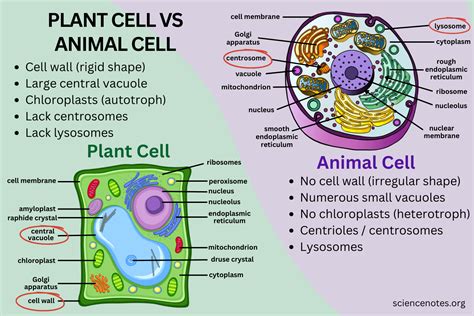
In conclusion, the animal cell diagram with labels is a valuable tool for understanding the intricate structures and functions of living organisms. Each organelle plays a unique role in maintaining cellular homeostasis, and together they work in harmony to sustain life.
What is the main function of the plasma membrane?
+
The main function of the plasma membrane is to regulate the movement of materials in and out of the cell.
What is the role of the mitochondria in an animal cell?
+
The mitochondria are responsible for generating energy for the cell through the process of cellular respiration.
What is the function of the Golgi apparatus in an animal cell?
+
The Golgi apparatus is responsible for protein modification, sorting, and packaging for transport out of the cell.
Related Terms:
- Animal cell structure
- Animal cell 3D
- Animal cell journal
- Animal and plant cell
- Plant cell structure
- Plants cells


